FIGURE 6.
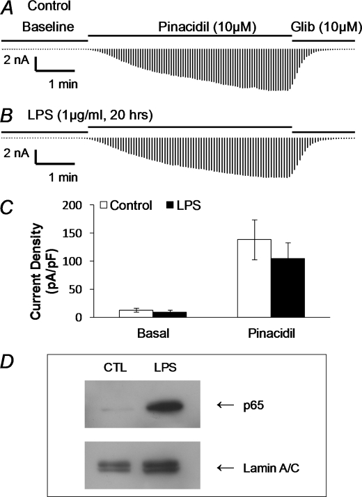
LPS failed to change Kir6.1/SUR2B channel activity in a heterologous expression system. A, Kir6.1/SUR2B were co-expressed with TLR4/MD2/CD14 in HEK293 cells, and whole cell currents were studied as shown in Fig. 2. The current amplitude increased markedly in response to pinacidil (10 μm), and was inhibited by glibenclamide (Glib, 10 μm). B, in another cell treated with LPS (1 μg/ml) overnight, the currents showed similar responses to pinacidil and Glib. C, comparison of the current density between the control (n = 15) and LPS-treated cells (n = 13). Both basal current density and pinacidil-induced current density were not significant changed after overnight LPS incubation (1 μg/ml, p > 0.05). D, stimulation of NF-κB signaling with LPS exposure in HEK293 cells. HEK293 cells were transfected with human TLR4/MD2/CD14 cDNAs. Two days after transfection, Western blot analysis was performed on the nuclear extracts from cells. The p65 accumulation was clearly seen 30 min after LPS stimulation.

