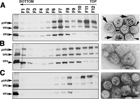
FIGURE 6.
VP3 C-terminal basic region is important for T = 13 capsid assembly. Western blot analysis of proteins expressed in cells infected with VT7/LacOI/R243D (A), VT7/LacOI/R246D (B), or VT7/LacOI/R249D rVV (C) is shown. Infected cultures were harvested at 72 h.p.i., and assemblies were purified by two-step centrifugation; 12 fractions were collected, concentrated, and analyzed by SDS-PAGE and Western blotting using anti-VP2 (top) or anti-VP3 (bottom) antibodies. The direction of sedimentation was right to left, with fraction 12 representing the gradient top. Images shown on the right correspond to representative electron micrographs (negative staining) of single-point polyprotein mutant assemblies: Poly/R243D T = 13 (arrows) and T = 7 (arrowheads) (A) capsids, Poly/R246D collapsed T = 13 capsid-like particles (B), and Poly/R249D T = 7 capsids (C). Scale bar, 100 nm.

