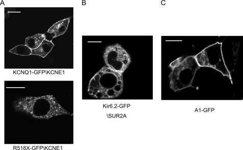
FIGURE 1.
Imaging with confocal microscopy in live HL-1 cells. A (upper), KCNQ1-GFP-KCNE1 localizes to the plasma membrane and (lower) the mutant KCNQ1(R518X)-GFP-KCNE1 shows diffuse punctate localization and weak localization to the plasma membrane. B, Kir6.2-GFP-SUR2A localizes partially to the cytoplasm. C, A1-GFP localizes to the plasma membrane. Scale bars are 10 μm.

