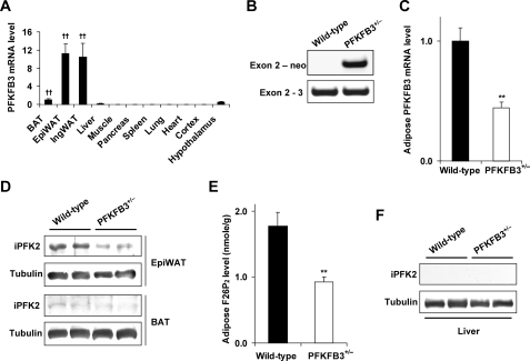
FIGURE 1.
Disruption of PFKFB3/iPFK2 decreases iPFK2 expression and activity in adipose tissue. A, wild-type C57BL/6J mice were used for analyses. PFKFB3 is abundantly expressed in epididymal (Epi) and inguinal (Ing) white adipose tissue. BAT, brown adipose tissue. Data are means ± S.E., n = 4. ††, p < 0.01 versus liver. B, PCR analyses of mouse genomic DNA using an exon 2-specific primer with a neomycin-specific primer (+/−, heterozygous) or an exon 3-specific primer (+/+, wild-type). For C and E, data are means ± S.E., n = 4 - 6. **, p < 0.01 PFKFB3+/− versus wild-type. C, levels of PFKFB3 mRNA in epididymal adipose tissue were quantified using real-time RT-PCR. D, amount of iPFK2 in both epididymal adipose tissue and brown adipose tissue was measured using Western blot. E, levels of F26P2 in epididymal adipose tissue were determined using the 6PFK1 activation method. F, amount of iPFK2 in the liver was determined using Western blot.

