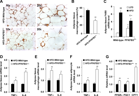
FIGURE 4.
Disruption of PFKFB3/iPFK2 exacerbates HFD-induced adipose tissue inflammatory response and increases adipose expression of genes involved in fatty acid oxidation. At the age of 5–6 weeks, PFKFB3+/− mice and wild-type littermates were fed an HFD for 12 weeks. A, macrophage infiltration in adipose tissue. The sections of epididymal fat pad were immunostained for F4/80. For B–G, data are means ± S.E., n = 4 - 6. *, p < 0.05 and **, p < 0.01 HFD-PFKFB3+/− versus HFD-wild-type (in B and D–G) or PFKFB3+/− versus wild-type on the same diet (in C); †, p < 0.05 and ††, p < 0.01 HFD versus LFD for the same genotype (in C). B, fraction of adipose tissue macrophages. C–G, mRNA levels of inflammatory markers and genes involved in fatty acid oxidation were quantified using real-time RT-PCR. C, mRNA levels of F4/80 in adipose tissue. D–F, mRNA levels of TNFα and IL-6 in adipose tissue (D), as well as in macrophages (E) and adipocytes (F) isolated from adipose tissue. G, mRNA levels of genes involved in fatty acid oxidation in adipose tissue.

