
FIGURE 1.
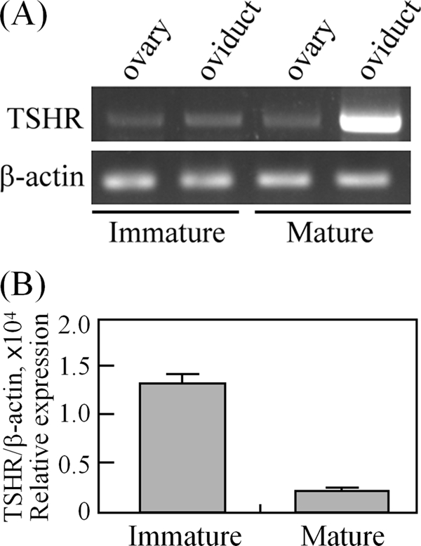
TSHR is expressed in the ovary and oviduct. cDNAs from immature (26 days old) and mature (8 weeks old) rat tissues were used for PCR amplification (A) and real-time quantification of TSHR expression levels (B). Data are expressed as the means ± S.D. The levels of β-actin served as loading and normalized controls.