FIGURE 5.
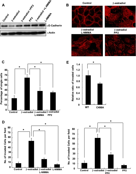
Disruption of cell-cell adhesion and induction of cell invasion after β-estradiol stimulation was mediated by NO and c-Src. A, MCF7 cells were stimulated with β-estradiol (100 nm) in the presence or absence of either L-NMMA (50 μm) or PP2 (25 μm) for 24 h, and expression of E-cadherin was examined by Western blot. B, MCF7 cells were stimulated with β-estradiol (100 nm) in the presence or absence of either L-NMMA (50 μm) or PP2 (25 μm) for 24 h and immunostained for E-cadherin. C, MCF7 cells were stimulated with β-estradiol (100 nm) in the presence or absence of either L-NMMA (50 μm) or PP2 (25 μm) for 24 h, and cell aggregation assay was performed. Percentages of single cells were counted, and mean averages + S.D. from three independent experiments are indicated. *, p < 0.01. D, MCF7 cells were loaded onto Matrigel-coated filters with or without β-estradiol (100 nm) in the presence or absence of either L-NMMA (50 μm) or PP2 (25 μm). Cells that invaded to the lower surface of the filters were fixed, stained, and quantified by counting three randomly selected fields under the microscope. Mean averages + S.D. from three independent experiments are indicated. *, p < 0.01. E, MCF7 cells were transfected with siRNA targeting 3′-UTR of human c-Src and a vector encoding green fluorescent protein together with either a vector encoding wild type avian c-Src or C498A Src. Two days after transfection, cells were subjected to the invasion assay in the presence of β-estradiol (100 nm). The number of cells that invaded to the lower surface was counted and normalized by transfection efficiency. Three independent experiments were performed, and the relative ratio of migrated cells is indicated. *, p < 0.01. Error bars indicate S.D.

