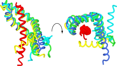
FIGURE 4.
Structural plasticity of Sus1. Worm traces of the different copies of the complex in the crystals superimposed to illustrate that although the relative positions of helices α2–5 were well conserved between different copies of the Sgf11-Sus1 complex, the position of helix α1 was very variable. The Sgf11 helix is shown in red; two chains from the complex with Sgf11 residues 7–33 are shown in blue and yellow, and two chains from the complex with Sgf11 residues 1–33 are shown in cyan and green.

