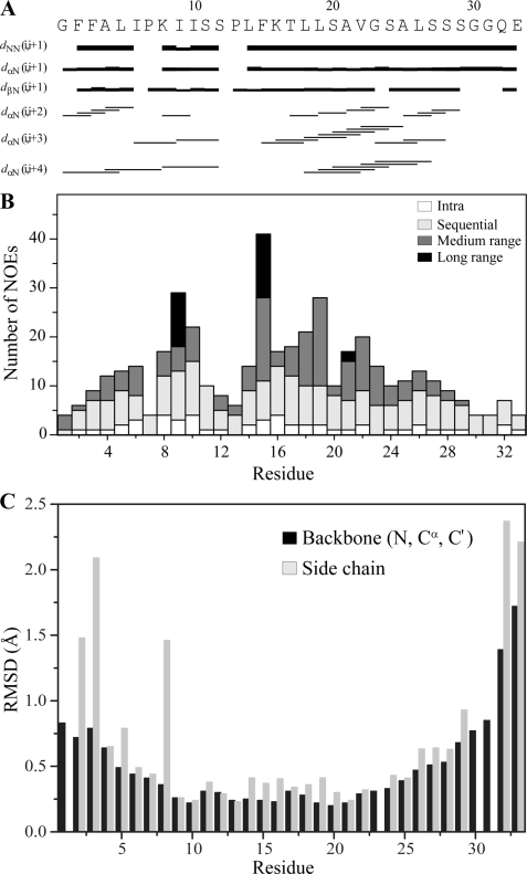
FIGURE 6.
A summary of NMR structural parameters of Pa4 in LPS micelle. A, a bar diagram showing sequential and medium range NOEs of Pa4 in the presence of LPS. The length of the bars indicates the intensity of the peaks, which are assigned as strong, medium, and weak. Amino acid sequence of Pa4 is shown at the top. B, a histogram showing the number of tr-NOEs of Pa4 as a function of residue number in complex with LPS micelles. C, root mean square deviations (RMSD) of backbone and side-chain atoms as a function of residue number of Pa4 for the twenty lowest energy conformers.

