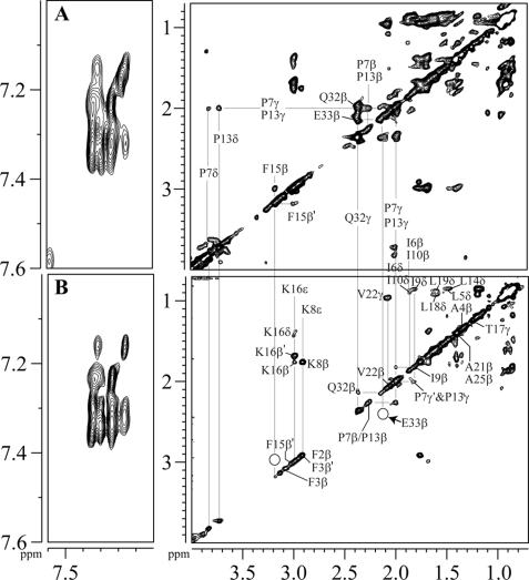
FIGURE 8.
Localization of Pa4 in LPS micelles by STD NMR. A, the reference or off-resonance TOCSY spectrum and (B) STD TOCSY spectrum of Pa4 in the presence of LPS in D2O at pH 4.5 and 288 K, showing the downfield-shifted aromatic resonances (left panels) and upfield-shifted side-chain resonances (right panels). Spectra were acquired using spin-lock MLEV17 sequence with a mixing time of 80 ms. Saturation of LPS was achieved by applying a cascade of 40 Gaussian pulses (50 ms each), resulting in a total saturation of ∼2 s (on-resonance, −2.0 ppm; off-resonance, 40 ppm).

