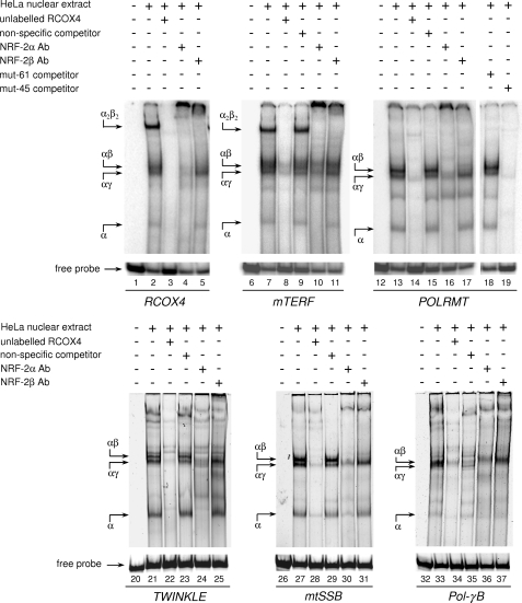
FIGURE 3.
In vitro binding of NRF-2 to the predicted sites in the promoter of the selected genes. NRF-2 binding was evaluated by EMSA using a heparin-Sepharose-purified nuclear extract from HeLa cells and radiolabeled double-stranded oligonucleotide probes containing one or two NRF-2 putative binding sites. The unlabeled specific (RCOX4), nonspecific, and mutated (mut-61 and mut-45) competitor oligonucleotides (100-fold molar excess) and NRF-2 antisera are indicated above each lane. Probes are shown below the lanes. DNA-protein complexes are indicated by arrows on the left of each panel. Ab, antibody.

