FIGURE 5.
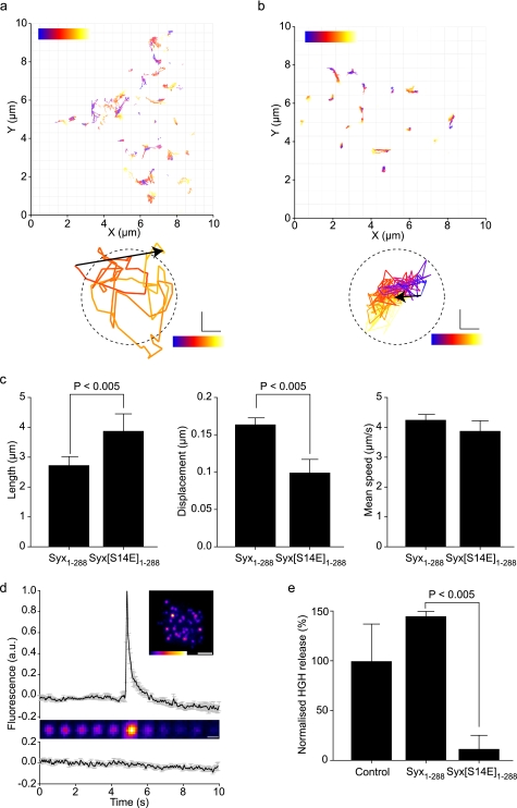
Vesicle behavior and fusion are controlled by the stability of the mode 2/3 interaction of syntaxin with Munc18. Fluorescent vesicles were tracked by TIRFM in cells expressing wild-type (a) or phosphomimetic (Syx1–288(S14E)) (b) syntaxin. Individual vesicle trajectories are shown as tracks with color corresponding to time during acquisition (blue, start; red, end). Examples most closely matching the mean track length and displacement for each condition are shown below. The dashed circle corresponds to mean vesicle diameter. Scale bar, 100 nm. c, individual tracks were measured for length, displacement, and mean speed (>250 tracks per cell; n = 4 cells). The phosphomimetic mutation resulted in enhanced track length but with a shorter displacement indicative of a tighter tethering. Mean speed was unaltered. Error bars are S.E. (t test, n = 4). d, cells expressing exogenous syntaxin 1 and Munc18-1 can perform exocytosis. The fusion of single secretory vesicles upon stimulation is shown as a fluorescence intensity trace and montage (upper panel; supplemental movie S3). Non-stimulated cells exhibited no detectable fusion events (lower trace). Error bars are S.E. (n = 15). e, exocytosis is severely depleted by disrupting the mode 2/3 syntaxin interaction with Munc18. The phosphomimetic form of syntaxin 1 (Syx1–288(S14E)) exhibited a pronounced inhibition of exocytosis. Error bars are S.E. (t test, n = 3).

