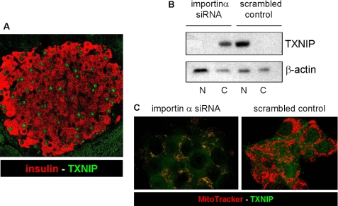
FIGURE 1.
Nuclear localization of TXNIP in pancreatic beta cells. A, immunofluorescent detection of endogenous TXNIP in wild-type C57BL/6 mouse pancreas sections by confocal microscopy. TXNIP was detected with the JY2 antibody and fluorescein isothiocyanate-labeled secondary antibody (green); beta cells were visualized by anti-insulin antibody and Cy3-conjugated secondary antibody (red). The pancreases of three mice were analyzed by staining of two separate 10-μm sections each; a representative pancreatic islet is shown. B, effects of importin-α1 siRNA knockdown on subcellular localization of TXNIP in INS-1 cells as assessed by cell fractionation and immunoblotting. N, nuclear fractions; C, cytoplasmic fractions. β-Actin was used as a loading control. One representative of three independent experiments is shown. C, confocal imaging of changes in subcellular localization of TXNIP in response to importin-α1 siRNA knockdown in INS-1 cells. Merged images were taken after 48 h: TXNIP (green) and MitoTracker Red (red)).

