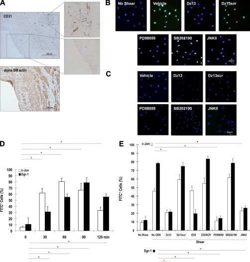
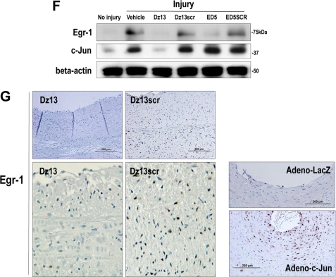
FIGURE 6.
c-Jun is induced in SMCs by fluid shear stress in an ERK1/2- and JNK-dependent and p38-independent manner and controls induction of the transcription factor Egr-1. A, CD31− α-SM actin+ cells reside at the inner surface of human saphenous vein conduits making contact with flowing blood (magnification, 200×). B, growth-quiescent SMCs, transfected with Dz13, Dz13scr, or incubated with PD98059, SB202190, or JNKII, were exposed to 10 dynes/cm2 fluid shear stress for 1 h and fixed, and immunocytochemical analysis was performed. c-Jun expression (FITC+) was visualized under fluorescence microscopy. Blue, DAPI-Blue staining indicates nuclei. C, SMCs were treated as in B, except that the cells remained under static conditions for the 1 h. D, SMCs were exposed to shear for the indicated times, and immunocytochemical analysis was performed for c-Jun or Egr-1 on fixed cells. FITC+ staining was expressed as a proportion of FITC+- and DAPI-Blue-stained nuclei in five random fields of view. E, effect of Dz13, Dz13scr, ED5, ED5 SCR, PD98059, SB202190, or JNKII on shear-inducible c-Jun and Egr-1 expression after 90-min exposure to fluid shear stress. FITC+ staining was expressed as a proportion of FITC+- and DAPI-Blue-stained nuclei in five random fields of view. F, Western blot analysis for Egr-1 or c-Jun using total cell extracts of SMCs (pretreated with Dz13, Dz13scr, ED5, or ED5SCR), injured by scraping and left for 1 h prior to harvest. In G: Left/upper panels, immunohistochemical analysis for Egr-1 in rabbit venoarterial autologous bypass transplants 28 days after the veins were treated ex vivo with Dz13, Dz13scr, and anastomosis. Right/lower panels, Egr-1 staining on vein transplants 28 days after the veins were transduced with Adeno-c-Jun or Adeno-LacZ.


