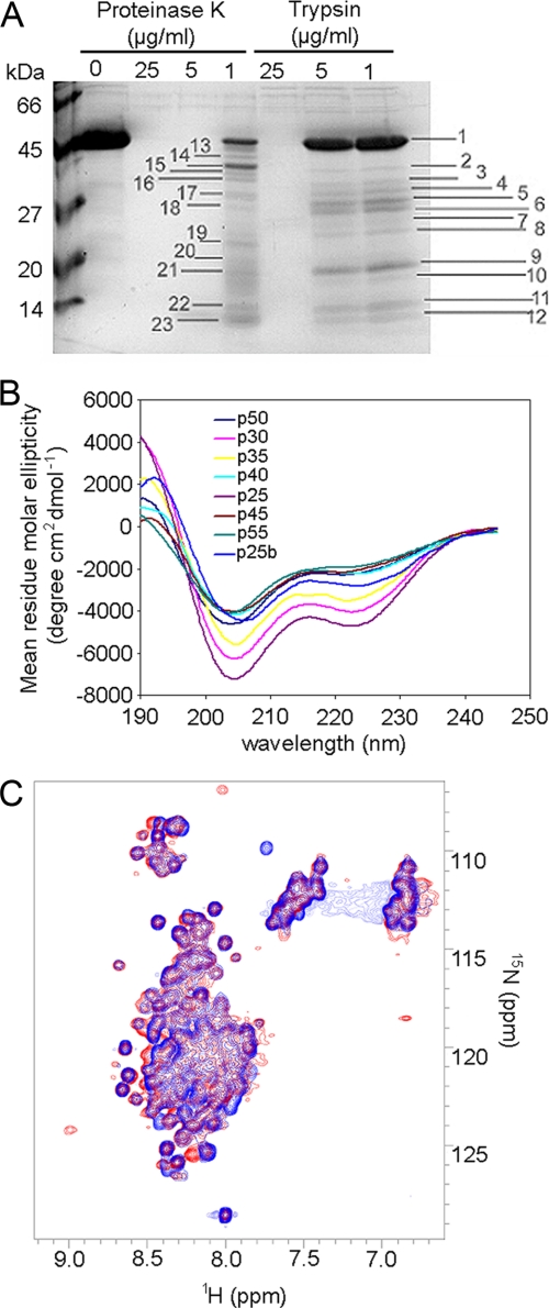
FIGURE 4.
Gadkin appears to lack stable tertiary structure. A, limited proteolysis performed with different concentrations of proteinase K and trypsin (1, 5, and 25 μg/ml) showed a pattern typical for a protein lacking stable tertiary structure. Fragments of different length were observed. Bands marked with numbers were in-gel-digested and analyzed by mass fingerprinting. All fragments could be assigned to the N-terminal portion of Gadkin (Δ51). B, CD spectra of 2.4 μm His6-tagged Gadkin (Δ51) in PBS, 10 mm dithiothreitol, taken at increasing temperature from 25 °C to 55 °C, shows the loss of structure elements with temperature increase to 40 °C. The process is irreversible; by cooling down to 25 °C (p25b) Gadkin (Δ51) remains partially unstructured. C, comparison of 1H,15N HSQC spectra of the 2H-, 15N-, and 13C-labeled Gadkin (Δ51) in the absence (blue) or presence (red) of γ-ear (1:2 ratio). Gadkin (Δ51) does not appear to adopt stable secondary structure upon addition of AP-1γ-ear.

