FIGURE 1.
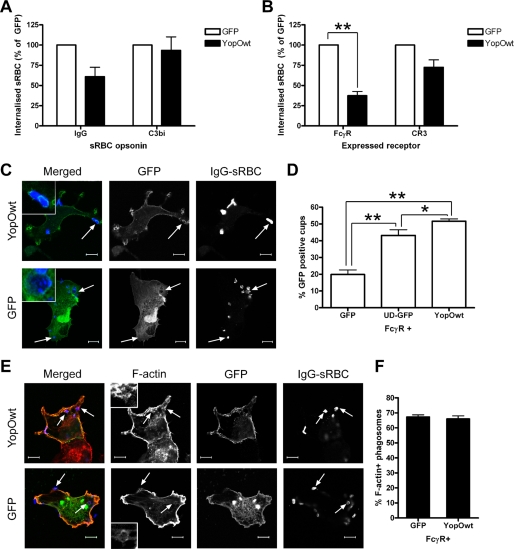
Preferential inhibition of FcγR-mediated phagocytosis by YopO. A, RAW264.7 mouse macrophages were transiently transfected with vectors expressing GFP or GFP-tagged YopOwt (YopOwt) and then challenged with IgG- or C3bi-opsonized sRBC for 30 min. B, COS-7 cells were co-transfected with FcγRIIA or CR3 vectors and either GFP or YopOwt and challenged with appropriately opsonized sRBC for 30 min. Phagocytosis assays were performed and scored as described under “Experimental Procedures.” At least 50 GFP-expressing RAW264.7 macrophages or 100 COS-7 cells were scored per experiment. Data represent mean ± S.E. from at least three independent experiments. C, COS-7 cells were transiently transfected with FcγRIIA and either GFP or GFP-YopOwt and then challenged with IgG-opsonized sRBC for 15 min after synchronization at 4 °C. sRBC and F-actin were labeled and observed by confocal microscopy. GFP enrichment at site of sRBC binding was examined. Note the localization of YopO, but not GFP around sRBC (arrows). D, quantification of GFP localization to phagocytic cups in cells co-expressing FcγRIIA and GFP, YopO-GFP, or unique domain of Lck-GFP (membrane-targeted GFP). E, following sRBC challenge, cells expressing GFP or YopOwt were analyzed for F-actin enrichment at sites of sRBC binding as described under “Experimental Procedures.” Representative images are shown, with typical F-actin rings enlarged. F, quantification of actin cup formation. At least 100 sRBC were scored for the presence of F-actin-rich cups per experiment. Data represent mean ± S.E. from at least three independent experiments. Scale bars, 10 μm. * indicates p < 0.05 and ** indicates p < 0.01.

