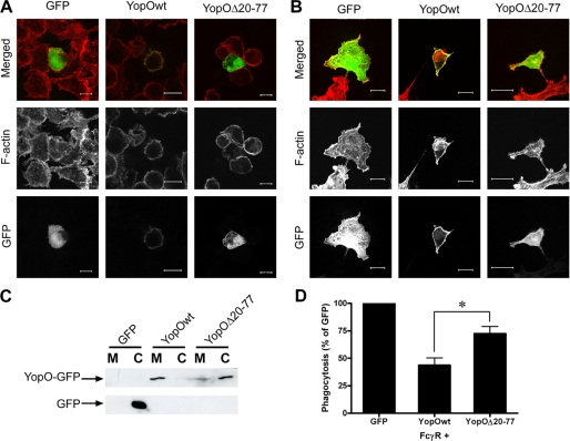
FIGURE 9.
Membrane localization of YopO is necessary for anti-phagocytosis. A, RAW264.7 macrophages were transiently transfected with GFP-tagged versions of YopO (wild type or Δ20–77) or GFP alone. After 24 h, cells were stained for F-actin and observed using a Zeiss Axiovert 100 M confocal microscope. Representative examples are shown. Scale bars, 10 μm. B, COS-7 cells were transfected and processed as described in A. Scale bars, 20 μm. C, lysates of COS-7 cells transfected with GFP-tagged constructs as indicated were separated into cytosol (C) and membrane (M) fractions, as described under “Experimental Procedures.” After SDS-PAGE, fractions were probed using anti-GFP antibodies. D, COS-7 cells were co-transfected with FcγRIIA and GFP-tagged YopO (wild type or Δ20–77) and challenged with IgG-sRBC, and at least 100 GFP-expressing COS-7 cells were scored for phagocytosis per experiment. Data are given as mean ± S.E. of at least three independent experiments. * indicates p < 0.05.

