FIGURE 1.
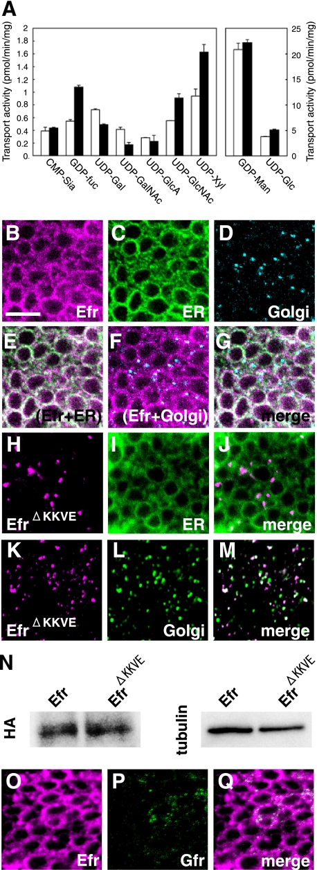
The Drosophila homolog of SLC35B4, Efr, transported GDP-fucose, UDP-N-acetylglucosamine, and UDP-xylose and was specifically localized to the ER. A, the transport of CMP-sialic acid (CMP-Sia), GDP-fucose (GDP-Fuc), UDP-Gal, UDP-GalNAc, UDP-GlcA, UDP-GlcNAc, UDP-xylose (UPD-Xyl), GDP-mannose (GDP-Man), and UDP-Glc into vesicles prepared from S. cerevisiae expressing Drosophila SLC35B4 from transfected pYEX-BX-Efr-C-HA (solid bars) or from the mock-transfected equivalent (open bars). Values are the mean ± S.E. from duplicate experiments. B–G, subcellular localization of HA-tagged Drosophila CG3774 (Efr-N-HA) in the epithelium of third instar wing discs. UAS-Efr-N-HA and UAS-CFP-ER were driven by ptc-Gal4. Efr (magenta in B, E, F, and G), CFP-ER (ER) (green in C, E, and G), and Golgi (cyan in D, F, and G) were detected by triple staining with anti-HA, anti-GFP, and anti-Golgi 120-kDa protein antibodies, respectively. E–G are merged images of B and C; B and D; and B, C, and D, respectively. H–M, subcellular localization of HA-tagged Drosophila CG3774 derivative lacking its dilysine ER retention/retrieval signals (EfrΔKKVE-N-HA) in the epithelium of third instar wing discs. UAS-EfrΔKKVE-N-HA and UAS-CFP-ER were driven by ptc-Gal4. EfrΔKKVE (magenta in H, J, K, and M) and CFP-ER (green in I and J) or Golgi (green in L and M) were detected by double staining with anti-HA and anti-GFP or anti-Golgi 120-kDa protein antibodies, respectively. J and M are merged images of H plus I and K plus L, respectively. N, a Western blot analysis of Efr-N-HA and EfrΔKKVE-N-HA proteins. UAS-Efr-N-HA and UAS-EfrΔKKVE-N-HA were driven by da-Gal4, and whole protein extracts were prepared from the embryos. These samples were analyzed by a Western blot using anti-HA antibody (HA). For an internal control, anti-β-tublin antibody (tublin) was used. O–Q, subcellular localization of Myc-tagged Drosophila CG3774 (Efr-N-Myc) and Gfr-C-HA in the epithelium of third instar wing discs. UAS-Efr-N-Myc and UAS-Gfr-C-HA were driven by ptc-Gal4, and Efr (magenta in O and Q) and Gfr (green in P and Q) were detected by double staining with anti-Myc and anti-HA antibodies, respectively. Q is a merged image of O and P. Bar, 5 μm.

