
Figure 1. Hyperglycemia increases NFATc3 nuclear accumulation in vivo.
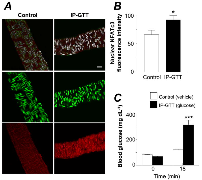
A. Confocal images showing NFATc3 nuclear accumulation in cerebral arteries from hyperglycemic (IP-GTT) and normoglycemic (control) C57Bl/6 mice. Upper panels are pseudo colored images showing nuclear NFATc3 in white; middle and lower panels are original images showing the DNA-binding dye SYTOX Green (green) and NFATc3 (red), respectively. Bar=20 μm. B. Summarized data from experiments in A showing fluorescence intensity of nuclear NFATc3 (N=6 in each group, 147 and 181 images per group, *P < 0.05). C. Glucose levels before and after IP-GTT, corresponding to experiments in A and B (***P < 0.001 vs. all groups).