Fig. 1.
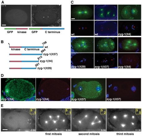
Elements within the C-terminus of ZYG-1 are important for localization to the centrosome and maintaining proper centrosome number. (A) GFP fused to the ZYG-1 C-terminus localizes to the centrosome (right), whereas ZYG-1 fused to the kinase domain is cytoplasmic (left). Scale bar: 10 μm. (B) Presumptive structure of the wild-type ZYG-1 protein and the three class II mutants. Due to mutation of a 3′ splice site, the zyg-1(it29) allele probaby produces a mixture of properly and improperly spliced messages. (C) Class II embryos stained for microtubules (green), DNA (blue) and centrosomes (red). Embryos are shown with (top) and without (bottom) microtubules. In the top set of panels, centrosomes are stained for SPD-2. The zyg-1(it4) embryo has failed to duplicate the sperm centrioles and as a result assembles monopolar spindles at the two-cell stage. In the bottom set of panels, the zyg-1(it29) embryo is stained for SPD-2, the zyg-1(it37) embryo is stained for SPD-5, and the zyg-1(it4) embryo is stained for SAS-4. Scale bar: 10 μm. (D) Young class II mutant embryos showing extra centrosomes exclusively associated with the male pronucleus. Centrosomes are stained for SPD-2. In the zyg-1(it4) embryo, the female meiotic spindle is visible (asterisk) but is not associated with centrosomes. In the zyg-1(it37) embryo, the female pronucleus (asterisk) lacks centrosomes. (E) Frames from a recording of zyg-1(it29) embryo expressing GFP-tubulin. Though the embryo inherits five centrosomes, they do not duplicate during the embryonic divisions. Numbers in yellow indicate the number of centrosomes (top) and cells (bottom) at each point in the recording. Time (minutes:seconds) is relative to first metaphase. Scale bar: 10 μm (D,E).

