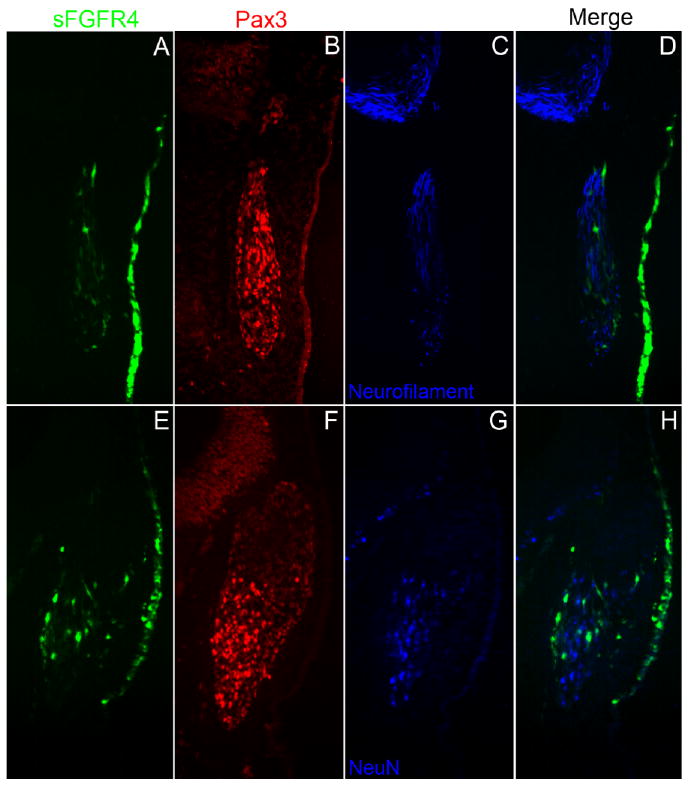
Fig. 5.

Secreted-fibroblast growth factor receptor-4 misexpression construct (sFGFR4) -targeted ophthalmic trigeminal (opV) placode cells do not differentiate as neurons in the ectoderm. A–C,E–G: Transverse section through the opV ganglion region of a ∼31 somite stage (ss) embryo, 36 hr after electroporation at the 7–9 ss with sFGFR4 (green; A,E), immunostained for Pax3 (red; B,F), Neurofilament (blue; C), and NeuN (blue, G). D: Merged image of sFGFR4 (green) and Neurofilament (blue). Cells in the ectoderm targeted with sFGFR4 do not up-regulate Neurofilament, although the neuronal marker is still expressed in untargeted cells in the opV ganglion. H: Merged image of sFGFR4 (green) and NeuN (blue); cells targeted with sFGFR4 do not delaminate and do not express the neuronal marker NeuN. Untargeted cells continue to migrate into the mesenchyme and express NeuN.
