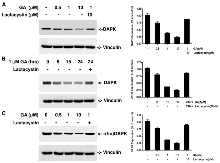
FIGURE 1. Geldanamycin treatment induces DAPK degradation through the ubiquitin proteasome pathway.
A, HeLa cells were treated with different concentrations of GA for 16 h in the presence or absence of lactacystin before lysis and analysis by Western blotting to detect the endogenous levels of human DAPK. Equal amounts of total cell protein were analyzed in each lane. B, HeLa cells were treated with 1 μM GA or 10 μM lactacystin for the indicated times and analyzed by Western blotting to detect the endogenous levels of human DAPK. C, HEK293 cells were transfected with 1 μg of pCDNA3-human DAPK. At 24 h, cells were then treated with Me2SO (vehicle) or the indicated concentrations of GA or 10 μM lactacystin for a further 16 h before analysis by Western blotting to detect recombinant human DAPK. Each blot is representative of three independent analyses, and vinculin is used as a loading control. r(hu)DAPK, recombinant human DAPK.

