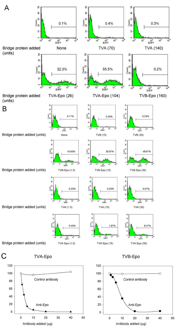
Figure 5.
Transduction of 293T-EpoR cells using ALV-A or ALV-B-pseudotyped lentiviral vectors preloaded with TVA-Epo or TVB-Epo bridge protein. (A) Specificity of TVA-Epo-mediated transduction. 293T-EpoR cells were transduced with ALV-A-pseudotyped LV-EGFP vectors preloaded with TVA-Epo or TVB-Epo bridge proteins and subjected to FACS analysis three days later. Aliquots of a concentrated vector stock corresponding to 5 × 10⁴ TU (determined on 293 DK-7 cells) were preincubated with cell culture supernatants containing TVA (upper center panel and upper right panel), TVA-Epo (lower left panel and lower center panel) or TVB-Epo (lower right panel). The amounts of bridge proteins added (expressed as V5 units) are indicated in parentheses. (B) Specificity of TVB-Epo-mediated transduction. 293T-EpoR cells were transduced with ALV-B-pseudotyped vectors preloaded with TVB-Epo and TVA-Epo bridge proteins and subjected to FACS analysis 3 days later. Aliquots of a concentrated vector stock corresponding to 1.2 × 10⁴ TU (determined on 293 DK-7 cells) were preincubated with cell supernatants containing TVB or TVB-Epo (top two panels), or TVA or TVA-Epo (bottom two rows). The amounts of TVB and TVB-Epo proteins added (expressed as V5 units) are indicated. Representative FACS profiles are shown. (C) Inhibition of TVA-Epo and TVB-Epo-mediated transduction of 293T-EpoR cells by anti-Epo antibody. 293T-EpoR cells were transduced using an EGFP-encoding lentiviral vector pseudotyped with the ALV-A Env (left panel) or the ALV-B Env (right panel). Vectors were exposed to cell supernatants containing TVA-Epo in the presence of monoclonal anti-Epo antibody or monoclonal anti-Flag antibody (as a control) on ice for 30 min. Transduced cells were subjected to FACS analysis three days later. The percentages of EGFP-positive cells are indicated. Representative data from two independent assays are shown.

