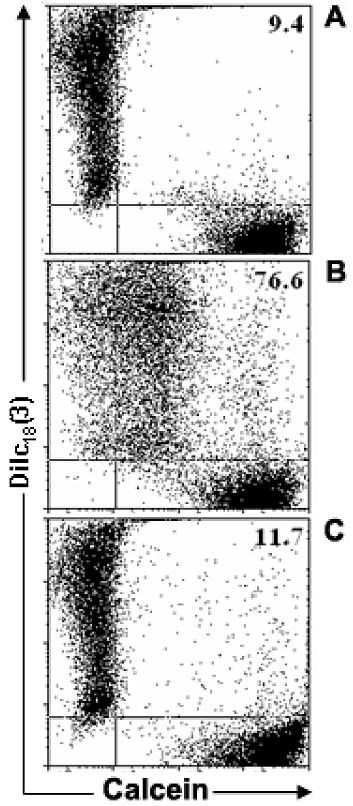
Figure 1.

Flow cytometric analysis of the mouse thymic epithelial cell line, showing inter-TEC gap junction intercellular communication. Calcein+Dilc18(3)- and calcein-DiIc18(3)+ IT-76M1 cells were co-cultured for 6 hr at 37°C. These cells were then dissociated and analyzed by flow cytometry to quantify the double positive [calcein+Dilc18(3)+] cells. Some calcein+Dilc18(3)- and calcein-DiIc18(3)+ cells were separately cultured and used to adjust the cytometry settings. These cells also were used to establish the control population (A). Data are presented in the form of dot plots (A, B, C), which depict two-dimensionally the labeling pattern of each cell population considering the fluorescence intensity (log scale) of calcein and DiIc18(3). In B, the 6 hr co-cultured cells are shown, where the presence of double positive cells is apparent, indicating the dye coupling. In C, cells co-cultured for 6 hours in the presence of 18-β-glycyrrhetinic acid (GRA; 100 μM) exhibited a complete inhibition of inter-TEC GJIC. These data are representative of at least 4 experiments.
