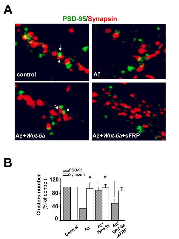
Figure 6.
Wnt-5a prevents the changes induced by Aβ oligomers in the synaptic contact.(A), Representative neurite images of double immunofluorescence of PSD-95 (green) and synapsin-1 (red), from samples subjected for 1 h to control, Aβ, Aβ/Wnt-5a and Aβ/Wnt-5a/sFRP treatments. Merged images show the apposition of the pre-synaptic (red) and post-synaptic (green) boutons. (B), Quantification of the number of clausters of the figure A (n = 3). Bar represents the mean ± SEM (*p < 0.05 Student's t test).

