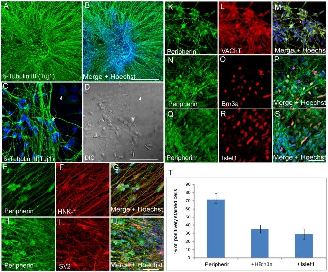
Figure 2. In vitro PNS differentiation capacity of hESC derived NPs.
(A–S) Eight weeks old Human NPs cultured on laminin for 12 days showing extensive neurite outgrowth expressing the pan neuronal marker β-tubulin III (A–D). These cells also express the PNS marker Peripherin (H,H,K,N and Q) together with the neural crest marker HNK-1 (F), the synaptic vesicle marker SV2 (I) and vesicular acetylcholine transporter VAChT (L), indicative of their maturity, the PNS markers Brn3a (O) and Istet-1 (R). (T) Quantitative analysis from these experiments (N–R) of the proportion of peripherin positive cells from total cells in the culture and the relative numbers of double stained Peripherin+/Brn3a+ and Peripherin+/Islet-1+ from total Peripherin+ cells. (Scale bar: in A-B = 0.5 mm; in C-S = 50 µm).

