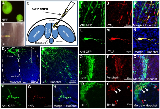
Figure 4. In vivo PNS differentiation of 8 weeks old hNPs implanted into the chick developing neural tube.
(A) Eight weeks old hESC expressing GFP derived NPs in suspension before cell dissociation. (B) Illustration of the cell transplantation procedure in the dorsal neural tube after removal of one neural fold. (C) Micrograph showing microinjection of GFP+ cells into the implantation site. (D–T) hNPs GFP+ derived cells 7 days after implantation located at the dorsal spinal cord showing extensive migration and neurite outgrowth (D and in high magnification in E). (F–H) implanted human NPs derived cells are specifically identified with anti-GFP antibodies (F) and with anti human specific nuclear antigen (G). Implanted hNPs express the human specific neuronal microtubule-associated protein Tau (I–K, and in high magnification in L–N). These cells also express the PNS markers, Peripherin (O–Q) and Brn3a (Q–S), Scale bars are indicated in representative images.

