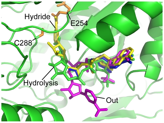
Figure 7. Different binding states of NAD(P)+ in the ALDH family.
Four different conformations of NAD(P)+ are shown as sticks, hydride conformation (PDB ID: 1bpw[31]: yellow), hydrolysis conformation (E. coli SSADH: green), out conformation (PDB ID: 2ilu [29]: magenta) and flexible, where the nicotinamide ribose moiety is unable to be resolved using X-ray crystallography (PDB ID: 2w8r [26]: blue). The general base (E254) and the catalytic cysteine (C288: both orange), which are conserved in human and E. coli SSADH and the whole ALDH family, have been labelled to define the active site.

