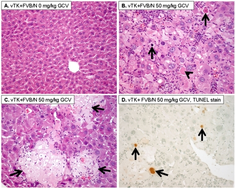
Figure 4. GCV induced histopathological changes.
A. vTK+FVB/N mice that did not receive GCV showed normal liver histology. B–C. vTK+FVB/N mice that received 50 mg/kg GCV showed increased cytoplasmic and nuclear enlargement with increased acidophilic bodies (B, arrowhead), apoptotic bodies (B, arrow), and areas of confluent necrosis (C, arrows) (A–C:Hematoxylin and eosin, original magnification ×200). D. TUNEL immunostaining showing increased apoptotic nuclei (arrows) in livers post GCV (Immunoperoxidase, original magnification ×200).

