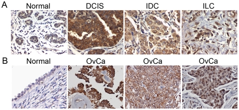
Figure 5. NPC expression is upregulated in breast and ovarian carcinomas.
(A) Immunohistochemical analysis of NPC expression was undertaken on a tumor tissue microarray (TMA) containing normal breast epithelial tissue and tumor specimens, including DCIS (ductal carcinoma in situ), IDC (infiltrating ductal carcinoma), and ILC (infiltrating lobular carcinoma). A mouse pan-anti-NPC monoclonal antibody was used for immunostaining. (B) Immunohistochemical staining of NPC on ovarian TMA containing three ovarian serous carcinomas (OvCa) compared to a normal ovarian surface epithelium (left panel). Images were taken at 400× magnification.

