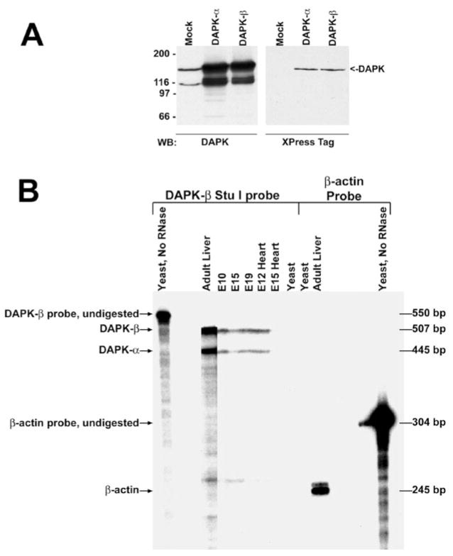
FIG. 2. Western blotting and ribonuclease protection to detect DAPK of mouse embryonic and adult tissues.
A, both panels are Western blots detecting expression of endogenous and recombinant DAPK-7α and DAPK-β in Cos cells. In the left panel, the recombinant DAPK detected in Cos cells transiently expressing DAPK-α or DAPK-β co-migrates with the endogenous DAPK detected in mock-transfected cells. In the right panel, an antibody to the Xpress tag detects the transiently expressed recombinant DAPKs. B, an autoradiogram of the ribonuclease protection analysis. The antisense cRNA probe is homologous to bp 4506–5009 of DAPK-β and contains 43 bp of vector sequence, making the total probe length 546 bp. Protection of a 507-and 445-bp probe fragment confirms that mRNAs corresponding to DAPK-β and DAPK-α, respectively, are present in adult liver and throughout development.

