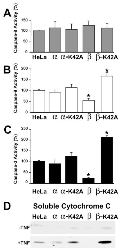
FIG. 8. Caspase activity and cytochrome c release in TNF-treated HeLa cell lines expressing wild-type and mutant DAPK.
For each experiment, at 24 h post-induction 10 ng/ml TNF and cyclohexamide (10 μg/ml) was added to initiate the experiment. Caspase-8 (A), caspase-3 (B), or caspase-9 (C) activity was determined directly in cell extracts by quantitating the cleavage of IETD-pNA (caspase-8), LEHD-pNA (caspase-9), or DEVD-pNA (caspase-3). The results shown represent the mean ± S.E. from at least three independent experiments. Significance (*) indicates that p ≤ 0.01. D, the release of cytochrome c from mitochondria was determined in the presence and absence of TNF by Western blotting of the post-mitochondrial supernatant from the indicated HeLa cell lines expressing DAPKs. The panel shown is representative of three independent analyses.

