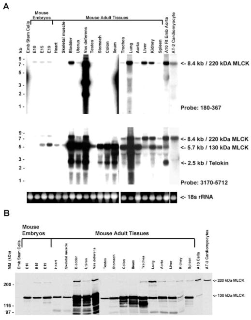
Fig. 3.
Northern and Western blotting to detect MLCKs. A: Northern blots in which 15 μg of total RNA, isolated from the indicated mouse tissues, was separated on a 1.4% agarose gel and probed with a cRNA probe corresponding to bp 180–367 (top) or bp 3170–5712 (bottom) of the mouse 220-kDa MLCK. Top, a single, 8-kb mRNA corresponding to the 220-kDa MLCK is detected. The smaller mRNAs detected in vas deferens are not reproducible and may represent genomic contamination. Bottom, 3 messages are detected: 8.4- and 5.7-kb mRNAs corresponding to the 220- and 130-kDa MLCKs and a 2.7-kb mRNA corresponding to telokin. Shown below the Northern blots is a photograph of 18s ribosomal RNAs to indicate the relative loading of the total RNA samples used in this analysis. B: Western blot analysis of extracts from various mouse tissues and cell lines, reacted with the K36 monoclonal antibody. A 130-kDa protein corresponding to smooth muscle MLCK is detected in most tissues and cells; a 220-kDa protein corresponding to nonmuscle MLCK is detected in several smooth muscle tissues and some cell lines. The lower-molecular-mass bands in some samples represent proteolytic degradation products.

