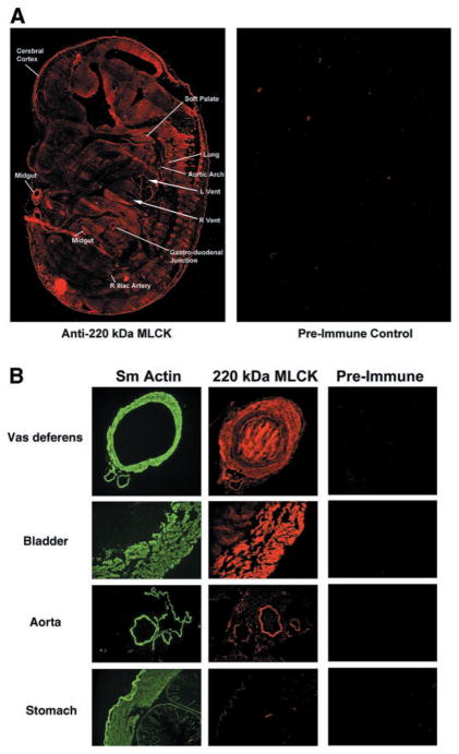
Fig. 4.
Localization of 220-kDa MLCK in mouse embryos and adult tissues. A: serial sections of mouse day 14.5 embryos were reacted with affinity-purified anti-220-kDa (1–170) antibody or preimmune control serum, followed by rhodamine-conjugated goat anti-rabbit IgG. The 220-kDa MLCK is detected in all tissues in the mouse day 14.5 embryo. The right and left ventricles of the heart (R vent, L vent) are indicated by arrows. The lines point to specific tissues that consistently had higher levels of expression of 220-kDa MLCK. B: localization of 220-kDa MLCK in adult tissues. Selected mouse adult tissues (vas deferens, bladder, aorta, and stomach) were fixed and reacted with antibodies to detect α-smooth muscle actin (fluorescence conjugated 1A4 antibody, Sm Actin), 220-kDa MLCK (1–170), or preimmune control serum followed by rhodamine-conjugated second antibody. Center panels, localization of 220-kDa MLCK with respect to the smooth muscle layers of the tissues detected in left panels with the α-smooth muscle actin antibody.

