
Fig. 5.
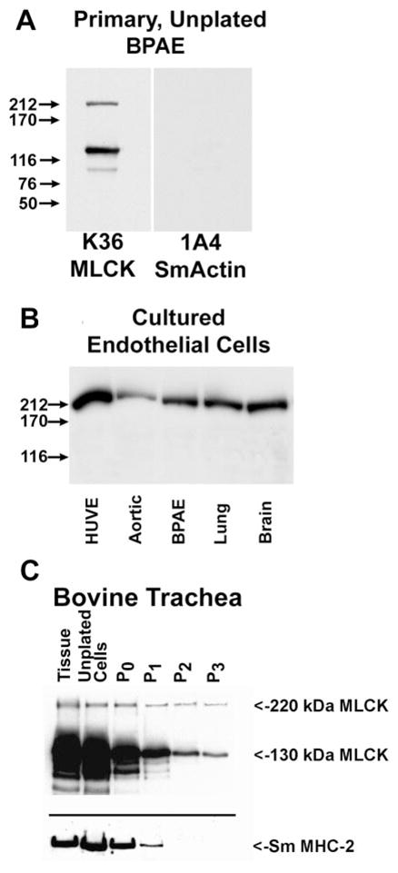
Changes in expression of MLCKs in cultured cells. Western blotting to detect MLCKs: primary uncultured bovine pulmonary artery (BPAE) cells (A), endothelial cells passed in culture (B), and bovine tracheal smooth muscle tissue and primary tracheal cells during passage in culture (C). HUVE, primary cultures of human umbilical vein endothelial cells. Aortic, BPAE, lung, and brain are all primary bovine endothelial cells that have been maintained in culture for several passages. In C, extracts of tracheal tissue (tissue), unpassaged freshly isolated tracheal smooth muscle cells, and cultured, passaged tracheal smooth muscle cells are compared. The passage (P) number for the bovine tracheal cells is indicated.