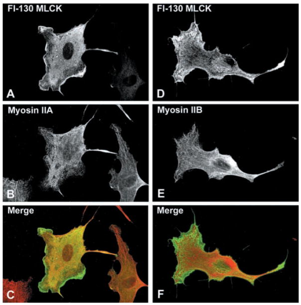
Fig. 7.
Intracellular localization of 130-kDa MLCK in BPAE cells. BPAE cells transiently expressing Fl-130-kDa MLCK were fixed and reacted with mouse anti-Flag antibody and Alexa 488-conjugated goat-anti-mouse secondary antibody to detect MLCK or with affinity-purified anti-NMHC-IIA or -IIB antibodies and Alexa 568-conjugated goat anti-rabbit IgG as indicated. Each vertical row represents the same cell expressing 130-kDa MLCK and merged images are shown in C and F (MLCK in green; NMHC-IIA or -IIB in red; overlap is yellow-orange). The staining pattern for the 130-kDa MLCK is predominantly cytoplasmic, although fine filaments are detectable that colocalize with NMHC-IIA but do not coincide with NMHC-IIB.

