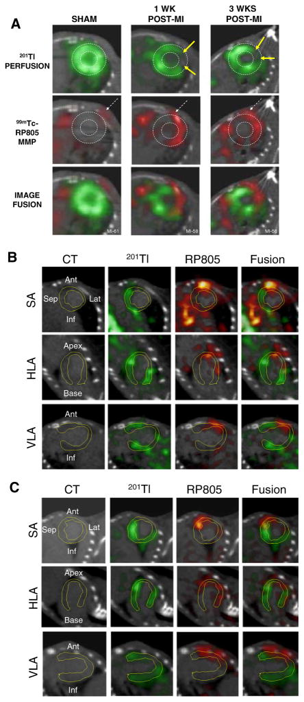
Figure 10.
Imaging of MMP activity postinfarction. Hybrid Micro-SPECT/CT reconstructed short-axis images were acquired without x-ray contrast (A) in control sham-operated mouse (left) and selected mice at 1 week (middle) and 3 weeks (right) after MI, after injection of 201Tl (top row, green) and 99mTc-RP805 (middle row, red). A black-and-white (B&W) and multicolor fusion image is shown on bottom. Control heart demonstrates normal myocardial perfusion and no focal 99mTc-RP805 uptake within the heart, although some uptake is seen in chest wall at the thoracotomy site (dashed arrows). All post-MI mice have a large anterolateral 201Tl perfusion defect (yellow arrows) and focal uptake of 99mTc-RP805 in defect area. A dashed circle is drawn around the heart to demonstrate localization of 99mTc-RP805, the MMP radiotracer, within the infarcted area of the heart. Some activity is also seen in the peri-infarct border zone. Additional micro-SPECT/CT images were acquired by use of a higher-resolution SPECT detector after the administration of x-ray contrast, at 1 week (B) and 3 weeks (C) after MI. The contrast agent permitted better definition of the LV myocardium, which is highlighted by white dotted line. Representative short-axis (SA), horizontal long-axis (HLA), and vertical long-axis (VLA) images are shown. Focal uptake of 99mTc-RP805 is seen within the central infarct and peri-infarct regions, which again corresponds to 201Tl perfusion defect (Reprinted with permission Su et al.101).

