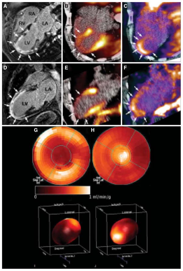
Figure 4.
Cine-MRI (CMR) with delayed enhancement (arrows) extending from the anterior wall to the apical region in the four-chamber (A) and two-chamber (D) view. (B, E) Identically reproduced location and geometry with severely reduced myocardial blood flow using 13N-ammonia, corresponding to the regions of delayed enhancement by CMR (arrows). (C, F) Focal 18F-RGD signal co-localized to the infarcted area. This signal may reflect angiogenesis within the healing area (arrows). (G, I) Polar map (I: 3D) of myocardial blood flow assessed by 13N-ammonia indicating severely reduced flow in the distal LAD-perfused region. (H, J) Co-localized 18F-RGD signal corresponding to the regions of severely reduced 13N-ammonia flow signal, reflecting the extent the of αvβ3 expression within the infarcted area (Reprinted with permission from Makowski et al.41).

