
Figure 7.
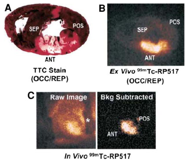
Imaging ischemic inflammation with the LTB4 receptor antagonist, RP517. TTC-stained heart slice (A) and ex vivo 99mTc-RP517 image (B) of the same heart slice. (C) Raw (left) and background subtracted (right) in vivo 99mTc-RP517 images acquired from a dog 60 minutes after reperfusion. Background subtraction was performed to eliminate the surgically related tracer uptake in the field of view. The shadow on the raw image denoted by an asterisk is the metal rib spreader. Note that focal 99mTc-RP517 uptake was readily observed in the inflamed anteroseptal region of the heart on both ex vivo and in vivo images. Tracer uptake was negligible in the normal, posterior wall. (Reprinted with permission from Riou et al.59).