
Figure 1.
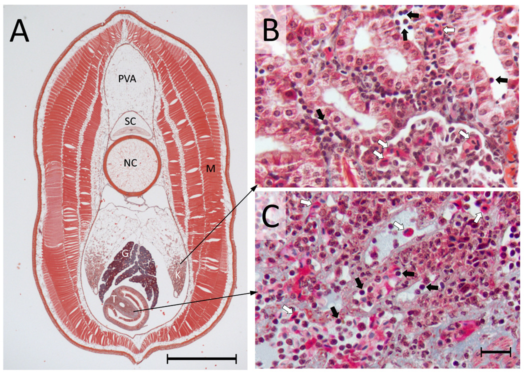
Distribution of cells in primary hematopoietic tissues in larval lamprey. (A) A transverse section from the mid-body region of an ammocoete lamprey (~ 13 cm in length). The 10 µm processed section stained with Masson Trichrome is showing different major internal organs, which include protovertebral arch (PVA), spinal cord (SC), notochord (NC), gonad (G), kidney (K), typhlosole (T) and muscle (M). Scale bar = 1 mm. (B) A magnified view of the kidney showing the distribution of blood cells, including many lymphocytes and erythrocytes (black and white arrows, respectively). Collections of blood cells are seen in and amongst the renal tubules. (C) A magnified view of the typhlosole showing diverse blood cells (lymphocytes and erythrocytes are indicated by black and white arrows, respectively). Scale bar = 10 µm. The structures that are stained light blue are largely extracellular matrix, which is highly abundant in the typhlosole.