
Figure 1.
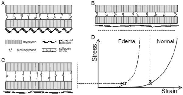
Contributions of ECM components to myocardial mechanics. A Schematic diagram shows myocytes connected by collagen struts, large perimysial collagen fibers aligned with the myocytes, and proteoglycans associated with the collagen. B During passive uniaxial stretch, titin in myocytes initially bears most of the force as perimysial collagen fibers uncoil; once straightened, collagen resists further extension, protecting myocytes from overstretch. C The location and mechanical role of cardiac proteoglycans are unknown; however, observed mechanical changes during edema could be explained by increased hydration of the proteoglycans pre-stressing the collagen network, as in cartilage. D The pre-stressed collagen network would resist deformation at much lower strains, shifting the uniaxial stress-strain curve to the left.