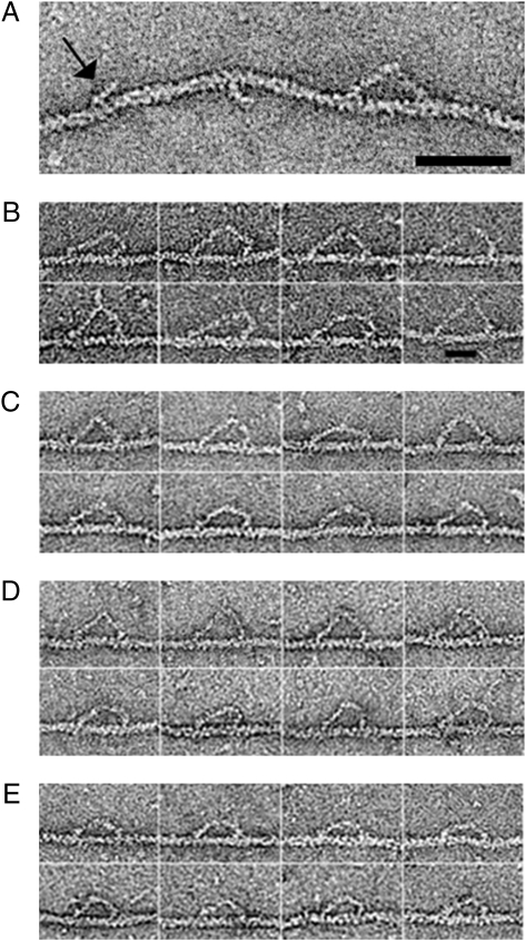
Fig. 1.
(A) Wild-type myosin 5a walking in ATP. Also attached are two single NEM-treated myosin 2 heads, one of which is indicated (arrow). These demonstrate the polarity of the actin filament. (Scale bar, 50 nm.) (B–E) Montages of wild-type and mutant myosin 5a HMM molecules bound to actin in 0.5 μM ATP, all walking to the right. (B) 8IQ, (C) wild type, (D) 2Ala-6IQ, (E) 4IQ. (Scale bar, 25 nm.)

