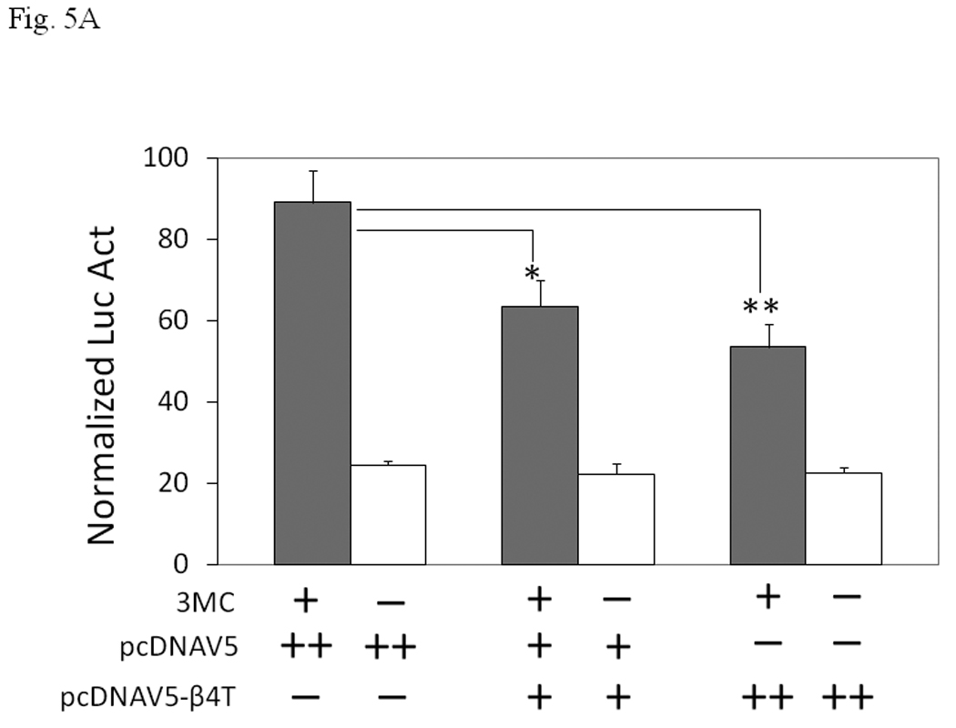
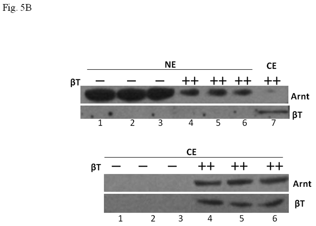
Fig. 5. Human β4-tubulin suppressed AhR signaling in Hep3B cells.


A. Transient transfection results showing suppression of the 3MC-driven luciferase activity by 250 ng (+) or 500 ng (++) of human β4-tubulin plasmid (pcDNAV5-β4T). Transfected DNA was normalized to 800 ng in all transfected samples with the empty plasmid pcDNAV5. Error bars show means ± SD (n = 3). *p < 0.01 whereas **p < 0.003. This experiment was repeated 4 times with similar results. B. Western blot analysis showing the effect of the transfected β4-tubulin on the nuclear and cytoplasmic Arnt content. Cells were transfected with 500 ng (−) of empty plasmid pcDNAV5 or 500 ng (++) of β4-tubulin plasmid pcDNAV5-β4T. Either 35 µg (top) or 50 µg (bottom) of proteins per lane was loaded. This experiment was performed in triplicate in the presence of 1 µM 3MC for 6 h: (Top) lanes 1–3, nuclear extract (NE) with empty plasmid; lanes 4–6, nuclear extract (NE) with β4 tubulin plasmid; lane 7, cytoplasmic extract (CE) with β4 tubulin plasmid. (Bottom) lanes 1–3, cytoplasmic extract (CE) with empty plasmid; lanes 4–6, cytoplasmic extract (CE) with β4 tubulin plasmid. Lane 7 (top) is the same sample as lane 4 (bottom), except that the film exposure time was different.
