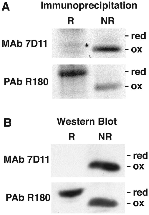
Fig. 1.
Specificities of L1 MAb and PAb for reduced and oxidized L1. (A) Immunoaffinity purification. BS-C-1 cells were infected with VACV and labeled from from 2 to 16 h with [35S]methionine/cysteine mixture. Lysates were prepared in the presence of DTT (R) or NEM (NR) and incubated with L1 MAb 7D11 or L1 PAb R180 complexed to protein G agarose. The proteins were eluted with sample buffer lacking reducing agent and analyzed by SDS-PAGE and autoradiography. Bands corresponding to reduced (red) and oxidized (ox) L1 are indicated. (B) Western blotting. Lysates were prepared as described in panel A and analyzed by SDS-PAGE and Western blotting with MAb 7D11 and PAb R180. Proteins were detected by chemiluminescence.

