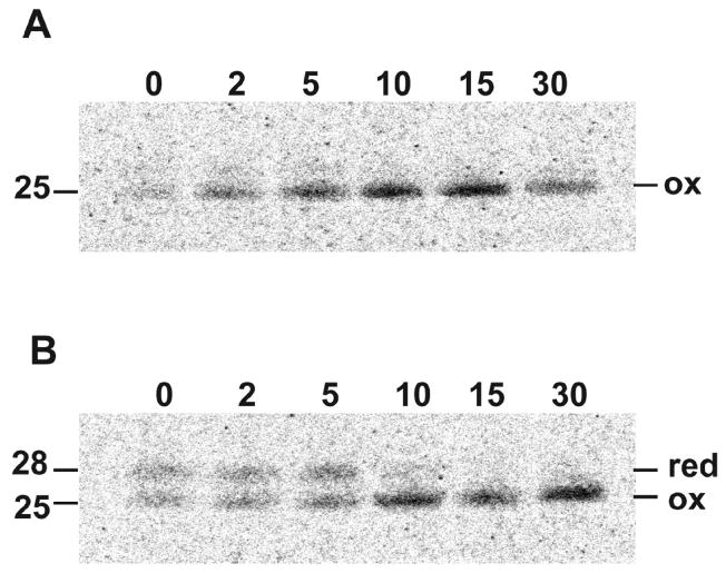
Fig. 2.
Kinetics of disulfide bond formation. BS-C-1 cells were infected with VACV for 16 h, incubated with methionine- and cysteine-free medium for 15 min, and then pulsed with 100 μCi of [35S]methionine/cysteine-labeling mix for 5 min. Following the pulse, cells were incubated in medium containing excess methionine and cysteine for 2 to 30 min as indicated, lysed, affinity purified with L1 MAb (A) or L1 PAB (B) and analyzed by SDS-PAGE and autoradiography. Bands corresponding to reduced (red) and oxidized (ox) L1 are indicated on the right. Numbers on the left indicate the masses of the L1 proteins in kDa determined by co-electrophoresis of marker proteins.

