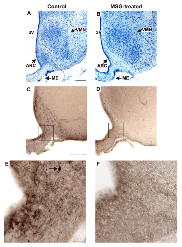
Figure 2.

Representative photomicrographs of adjacent sections in the mid-level of the arcuate nucleus in control (A, C, E) and MSG-treated (B, D, F) rats. (A, B), Methylene blue stain; (C, D, E, F), NKB-immunoreactivity with nickel intensified DAB. The boxes in C and D show the region illustrated in the high magnification photomicrographs of E and F. The Nissl stains from the MSG-treated rats revealed marked degeneration of neurons at most levels of the arcuate nucleus. Immunocytochemical stains confirmed the near-total loss of NKB-ir cells in the arcuate nucleus of MSG-treated animals. The high magnification photomicrographs illustrate loss of cell bodies and fibers in the arcuate nucleus and adjacent median eminence. The arrow in E points to an NKB-ir somata. Scale bar in A = 250 μm (applies to A and B), Scale bar in C = 200 μm (applies to C and D). Scale bar in E = 25 μm (applies to A-B).
