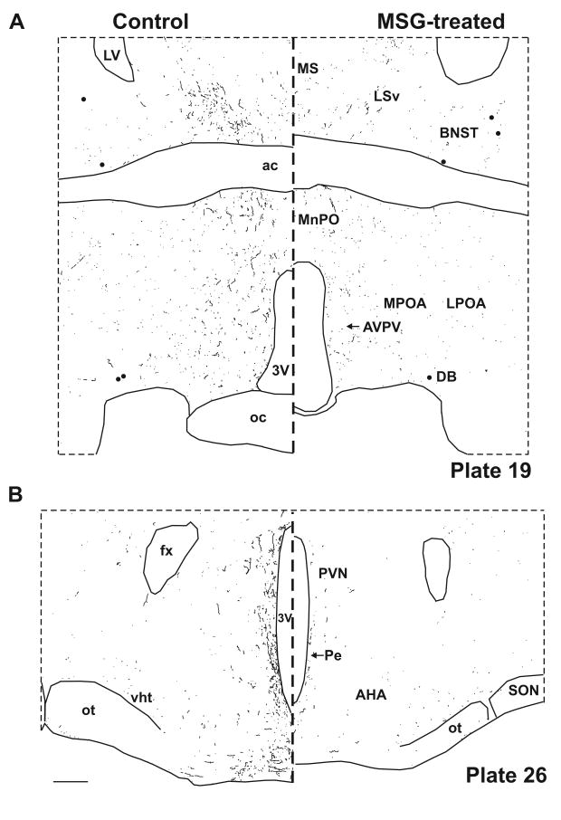
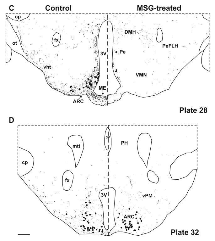
Figure 4.
Computer assisted drawings of NKB-ir neurons (filed circles) and fibers (lines) in control (left) and MSG-treated (right) rats at selected levels of the hypothalamus (A-D). Sections were matched to plates in a rat brain atlas listed in bottom right corner (Swanson, 1992). NKB-ir cells are depleted at most levels of the arcuate nucleus, with near total loss of NKB-ir fibers in the arcuate nucleus and the adjacent median eminence (C). NKB-ir fibers are also reduced in multiple hypothalamic areas (A-D). There is relative preservation of NKB-ir neurons at the level of the premammillary arcuate (D). Scale bars in B and D = 250 μm (applies to all).


