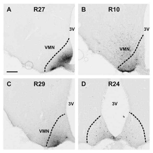
Figure 6.

Photomicrographs of BDA injection sites visualized by avidin-biotin-horseradish peroxidase histochemistry (A-D). The dotted lines indicate the border of the arcuate nucleus. The animal identification number is listed at the top of each photomicrograph. BDA injections were either confined to the arcuate nucleus (A, C, D) or included the arcuate nucleus and adjacent ventromedial nucleus (B). In one animal (D), the injection was midline and BDA was taken up in neurons in on both sides. Two injections (A and B) were at the mid-level of the arcuate nucleus and two (C and D) were in the posterior arcuate nucleus, corresponding to plates 28 (-2.45 from Bregma) and 31 (-3.70 from Bregma), respectively (Swanson, 1992). Scale bar in A = 200 m (applies to all).
