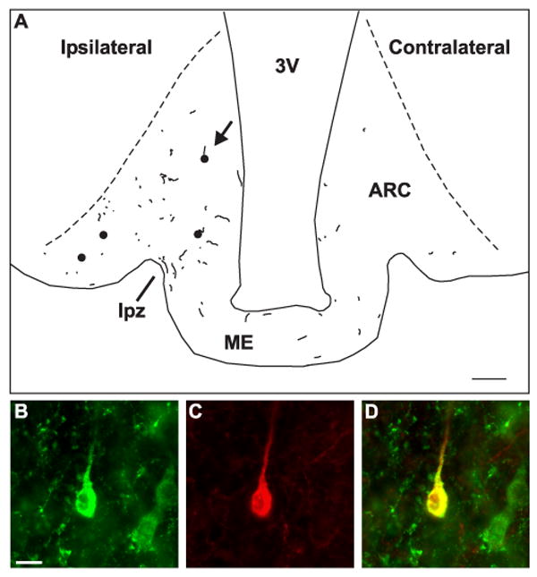
Figure 7.

A) Computer-assisted drawing showing the location of dual-labeled NKB/BDA-ir cell bodies (filled circles) and fibers (lines) near the injection site of R10 (see Fig. 6). This illustration was created by superimposing the tracings from 4 non-adjacent sections. The dotted lines indicate the borders of the arcuate nucleus. The arrow points to the location of the cell illustrated in B-D. Injection of BDA into the arcuate nucleus labeled a small number of NKB-ir neurons in the arcuate nucleus. Antereogradely labeled NKB-ir fibers were identified within the arcuate nucleus and adjacent median eminence including the lateral palisade zone (line). In addition, NKB/BDA-ir fibers were identified across the ME and within the arcuate nucleus of the contralateral side. (B-D) Demonstration of uptake of BDA in a NKB-ir neuron in the arcuate nucleus. High magnification photomicrographs of a NKB-ir cell body (B, green) in the arcuate nucleus that has taken up BDA (C, red). Color-combined image showing co-localization of NKB immunoreactivity and BDA (D, yellow). Scale bar in A = 100 μm. Scale bar in B = 10 μm (applies to B-D).
