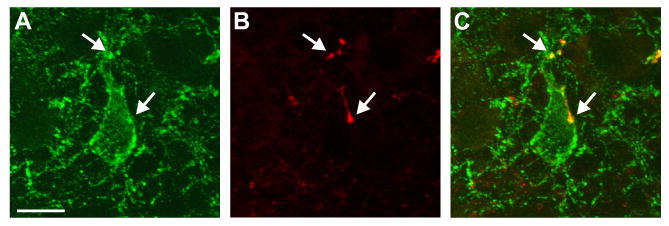
Figure 8.

Confocal microscope projection image of close apposition of a NKB/BDA-ir fiber onto a NKB-ir cell in the arcuate nucleus. NKB immunoreactivity is green (A) and BDA is red (B). The arrows point to dual-labeled fibers (yellow) in close apposition to the somata and proximal dendrite of an NKB-ir neuron ipsilateral to the side of injection. The projection is a stack of seven 0.8 μm thick confocal images. Scale bar in A = 10 μm (applies to all).
